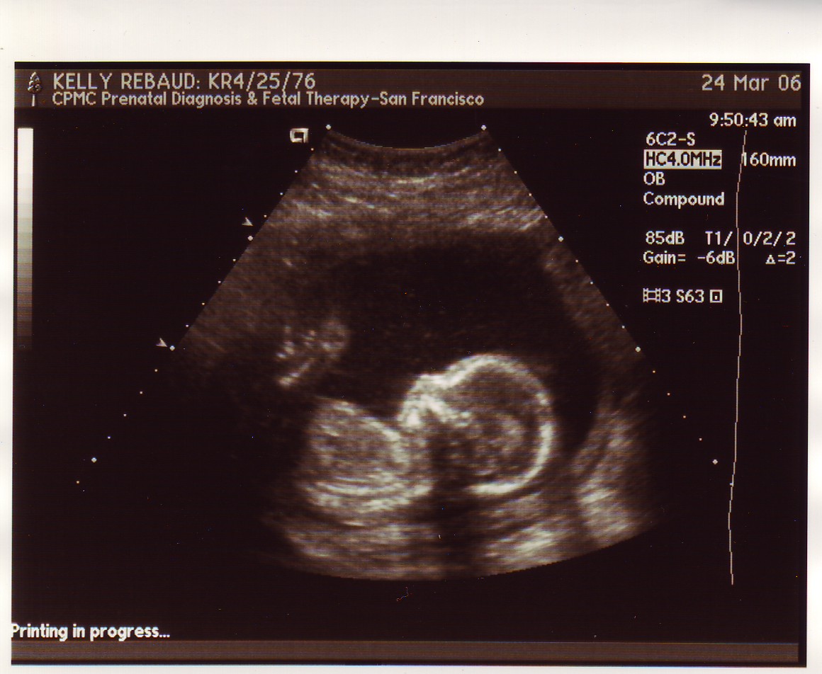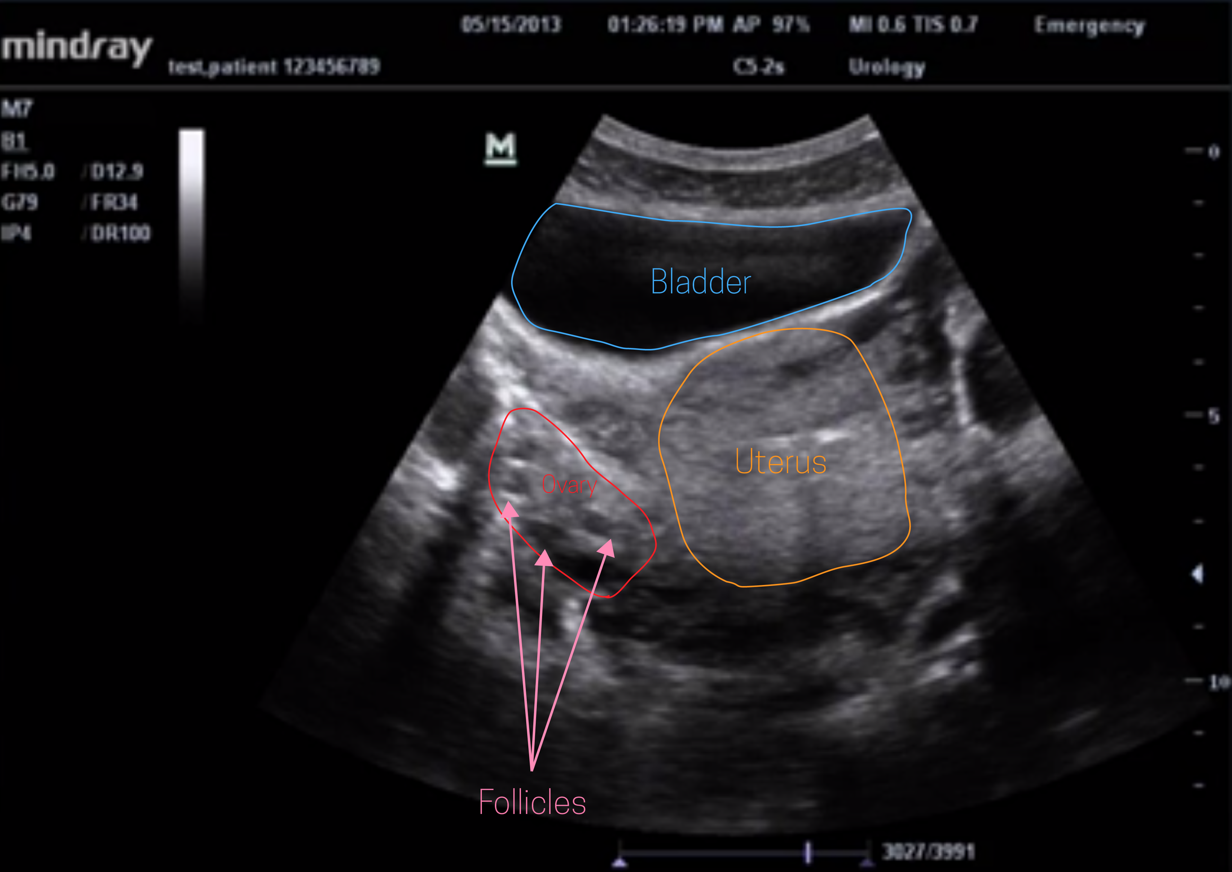

Sonograms are typically black and white, with solid whites representing hard tissues (such as bones), grays representing soft tissues (such as tendons and muscles), and blacks representing fluid (for instance, the fluid found in your amniotic sac). The Adelaide Women’s Imaging doctor will discuss the results of the scan with you on the day in the ultrasound room. Sonogram A sonogram is the visual image produced by the ultrasound procedure. This improves the accuracy of diagnosis and greater detail may be obtained of the fetal anatomy. It is disinfected, and a protective cover is placed over the transducer each time it is used, so there is no risk of infection. The transducer is lubricated with gel before insertion into the vagina. Transvaginal ultrasound involves inserting a thin transducer (ultrasound probe slightly thicker than a tampon) into the vagina to get more detailed images as the transducer is closer to the pelvic organs. Occasionally, the scan may need to be performed through the vagina (transvaginal imaging). The sonographer will perform an ultrasound of the abdomen (transabdominal ultrasound) detailing the fetal anatomy and maternal pelvic anatomy. The anomaly scan, also sometimes called the anatomy scan, 20-week ultrasound, or level 2 ultrasound, evaluates anatomic structures of the fetus, placenta. A sonographer will then collect you from the waiting room and take you to the ultrasound room. When you visit Adelaide Women’s Imaging for a 13 week anatomy scan you will be greeted by one of our friendly reception staff.
ANATOMY SONOGRAM FULL
Then slowly drink 600ml of water to fill your bladder and keep it full for your examination. Please empty your bladder 2 hours before the examination time. Two-piece clothing is ideal (separate upper/lower garments). If possible, please wear comfortable, loose-fitting clothing that allows easy access to the area that is being imaged.

Ideally, please call to book your appointment as early in the pregnancy as possible, so that the procedure can be done during the suggested period. Transabdominal and transvaginal ultrasound examinations are safe at all stages of pregnancy.

In most cases the scan will be performed transabdominally, but there are some situations where a transvaginal (internal) ultrasound may be necessary. Transrectal ultrasound, sometimes used in the diagnosis of prostate conditions, uses a transducer wand that is placed in the rectum.The 13 week anatomy scan is often referred for when a Non-Invasive Prenatal Testing (NIPT) blood test, commonly known as the Harmony Prenatal Test or NEST, has been undertaken for first trimester screening. If both babies 'cooperate' meaning they are able to get all the measurements and see everything as babies are in good positions and the ultrasound tech is experienced it shouldnt take longer then an hour. The anatomy scan, a thorough scan of your babys developing body and organs, is offered to every pregnant person.Transvaginal ultrasound uses a transducer wand that is placed in a woman’s vagina to get images of her uterus and ovaries.At times, a better diagnostic image can be generated with the insertion of a special transducer into one of the body’s natural openings: Most ultrasounds are done using a transducer on the surface of the skin. High intensity focused ultrasound (HIFU) has been designed to destroy or modify abnormal tissue in the body without opening the skin.Therapeutic ultrasound is used to heat or break up tissue.Bone sonography is used to determine bone density.Elastography is used to differentiate tumors from healthy tissue.But aside from the two standard ultrasounds, your doctor may schedule additional screenings to. Doppler ultrasound can be used to measure and visualize blood flow in the heart and blood vessels. This ultrasound checks for overall fetal development and is sometimes called the anatomy scan.These echoes generate electrical signals that are translated by a computer to produce images of the tissues and organs. Ultrasounds use high-frequency sound waves that are beamed into the body and bounce back (echo) off tissue and organs.


 0 kommentar(er)
